Huma oral cavity is highly complex, and several muscles work in sync to make it perfectly functional to help you eat, speak, and smile with ease. One such group of the membranous fold of mucous membrane and connective tissue is the frenum. This thin line runs between the gums and lips and the underside of the tongue to provide stability to the upper and lower lips and the tongue. The simple-looking anatomic structure, mostly ignored by most dentists during the regular examination, could be a sign of some syndromes. The shape, thickness, and length of the frenum vary among people of different ethnic groups.
Most people have perfect frenum attachment, but it can get snagged or pulled by overstretching or injury while eating, kissing, or wearing orthodontic braces. It bleeds heavily but heals naturally with some medication. If your frenulum is attached too close to the gingival margin, it could cause gingival health conditions as it interferes with brushing resulting in muscle pull. In such cases, your dentist might recommend the removal of the frenum by a special surgical procedure called a frenectomy.
Types of Frenum
The shape and length of frenum attachments change with the growth of the alveolar process along with the growth of the teeth. Your pediatrician will examine the frenum to detect any abnormality, as this could be a symptom of some serious ailments. Anatomically, there are two types of a frenum in the human oral cavity. These are:
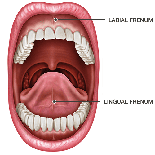
Lingual Frenum
The thin fold of mucous membrane with enclosed muscle fibers that connect the base of the tongue to the floor of the mouth is called the lingual frenum. It controls the movement of the tongue, so if it is tight it could affect the infant’s nourishment and cause speech difficulty in the future. If it is within the manageable range, your doctor will advise you to wait till teeth start erupting. If it is too tight, your doctor might recommend a frenectomy.
Labial Frenum
The dense collagenous tissue and elastic fibers between the lip and gum in the front of the mouth are called the labial frenum. There are two types of labial frenums, namely maxillary labial frenum and mandibular labial frenum.
The soft tissue connecting attaching the upper lip to the upper gum is called the maxillary labial frenum. This fold, also known as the superior labial frenum, originates at the midline of the undersurface of the lip.
If the shape and length are abnormal, it could affect the growth of teeth as it pulls gums away from the tooth root.
The soft tissue attaching the lower lip between mandibular central incisors with gingiva is called the mandibular labial frenum. If the attachment is abnormal it could cause gingival recession. Your doctor might recommend gingival reconstruction.
Frenum Abnormalities and Associated Conditions
The main functionality of the frenum is to provide stability to lips and tongue. If there is an anatomical abnormality or injury in the frenum, it could affect several associated functions of the oral cavity. If there is abnormal frenum development you might experience these conditions:
- Difficulty in swallowing
- Irregular tongue and lips movement leading to speech impairment
- Misaligned teeth development
- Gaps in front teeth
- Tearing of frenulum
- Infants might face nursing difficulty
- Abnormal jaw development casing snoring
- Gum injuries and infections
- Gum recession and teeth root exposure
Syndromes
Some of the syndromes associated with abnormal frenal attachments are:
Oro-facial-digital syndrome: This syndrome arises due to a single gene malformation showing X-linked dominant inheritance. Some of the clinical features of this syndrome are lobulated tongue with incompletely differentiated lingual frenum, cleft gums, cleft in the soft palate, or midline notch.
Infantile hypertrophic pyloric stenosis: The absence of mandibular frenum could be the symptom of this syndrome. Although the reason is unknown, it is more common in males.
Holoprosencephaly: If the labial maxillary frenum is absent or abnormal, it could be the symptom of holoprosencephaly. This autosomal dominant condition is characterized by brain malfunction.
Ellis-van Creveld syndrome: This is an autosomal recessive disorder that affects enamel, nail, and hair. Abnormal frenum attachment and the congenitally missing tooth could be associated characteristics of this syndrome.
Ehlers-Danlos syndrome: The absence of inferior labial and lingual frenum could be Ehlers-Danlos syndrome. People with this genetic disorder experience hyper extensive skin and hyper-mobile joints.
Pallister-hall syndrome: Abnormal supernumerary frena extending from the buccal mucosa to the alveolar ridge could be the sign of this genetic disorder. A short mid-face and nose with a flat nasal bridge are some of the features of this syndrome.
Opitz C syndrome: This syndrome also shows similar frenal attachment abnormalities as in Pallister-Hall syndrome.
What is Frenectomy?
The shape, position, and length of the frenum give strong cues about hidden genetic abnormalities. If you notice any abnormal frenum attachment growth in your infants and kids, you should consult a specialist before opting for its removal. Following a detailed examination, your doctor might recommend the removal of the frenum to improve oral functionalities using an especially designed surgical procedure called a frenectomy.
The procedure involves reduction of frenum length of tightness to remove undesirable effects of abnormal frenum growth. Although frenectomy is a simple surgery, doctors recommend this procedure only when it disrupts the normal functioning and growth of the mouth or tears repeatedly. If the abnormal frenum is affecting your kid’s speech ability or your infant’s nursing, then you might have to opt for frenectomy for your kid.
Depending on the abnormality of the frenum and associated structures like teeth, gums, and jaws, your doctor might use any of these three surgical techniques to remove frenal attachment:
Simple excision technique
If the fibrous and mucosal tissue band is narrow, your surgeon might use this surgical technique. He will make a narrow elliptic incision around the frenal area and then the fibrous frenum is dissected from the underlying soft tissues.
Z-plasty technique
For narrow frenum attachments, a surgeon uses this surgical technique. First fibrous tissue is removed and then two oblique Z-shaped incisions are made, one at each end of the previous area of excision. The flaps are then rotated to close the initial vertical incision.
Localized vestibuloplasty
If the frenal attachment has a wide base, surgeons opt for localized vestibuloplasty. In this procedure, a surgeon makes an incision through mucosal and submucosal tissues without piercing the periosteum. Then the supraperiosteal is dissected to identify a clean periosteal layer. The edge of the mucosal flap is sutured to the periosteum at the maximal depth of the vestibule.
What to Expect During a Frenectomy?
After a thorough examination, your oral surgeon will administer local anesthesia before performing frenectomy surgery. In most cases the procedure is short, and it takes just a few days to restore normal oral functionalities.
Depending on the complexity and surgical procedure opted for the procedure is performed using a scalpel via electrosurgery or with lasers. The use of sedatives depends on the age and comfort of the patient and the complexity of the frenal attachment. In simple cases, local numbing is enough, but if required your doctor might administer general anesthesia. You will remain unconscious for a few hours and won’t feel any pain during the procedure.
Your oral surgeon will perform the procedure to remove mucosal tissues of the area and suture the opening. Your doctor will prescribe some non-steroidal and anti-inflammatory medications to ease pain and make recovery easy. He will suggest some oral care measures and preventions to help you regain fully functional oral space faster.
How much does Frenectomy surgery cost?
The cost of the frenectomy, a surgical procedure to remove mucosal soft tissues of the mouth, averages between $500 and $1,500. However, the cost could vary widely depending on the complexity, the surgical procedure used, location of the clinic, and of course the experience of your oral surgeon. The type of sedative used could be an additional factor. You should get in touch with your health insurance provider to know about frenectomy coverage.
Bottom Line
The frenum is very important for oral functionality, as it offers stability to lips and tongue to ensure easy swallowing and smooth speech. The functional aspect is the same in all humans, but the shape and size of the frenum vary from person to person. Excessive stretching of the tongue and lips could tear frenum attachments, resulting in bleeding and pain. Tearing of the frenulum is very common, and it is not a matter of concern in most cases. However, if abnormal growth of the frenum affects swallowing, speech, or any other oral functions, your doctor might recommend surgical removal of frenum attachment.
Since frenulum abnormalities are linked with genetic disorders, early diagnosis and treatment could be of great help in ensuring the healthy development of your kids. So, it is always better to consult your dentist regularly so that he could monitor the growth of the frenum. Simple surgical intervention could prevent teeth misalignment, gaps, and bite issues. Ignoring isn’t a good option as tearing of frenulum regularly will affect your oral health, leading to some serious gum infections. Get in touch with your dentist to keep your mouth healthy.
Sources:
- Henry, S. W., Levin, M. P., & Tsaknis, P. J. (1976). Histologic features of the superior labial frenum.
https://europepmc.org/article/med/1063851 - Priyanka, M., Sruthi, R., Ramakrishnan, T., Emmadi, P., & Ambalavanan, N. (2013). An overview of frenal attachments.
https://www.ncbi.nlm.nih.gov/pmc/articles/PMC3636930/ - Edwards, J. G. (1977). The diastema, the frenum, the frenectomy: a clinical study.
https://europepmc.org/article/med/266363 - Devishree, S. K. G., & Shubhashini, P. V. (2012). Frenectomy: a review with the reports of surgical techniques.
https://www.ncbi.nlm.nih.gov/pmc/articles/PMC3527809/